Page 85 - EASL POSTGRADUATE COURSE
P. 85
of obesity and its metabolic consequences, including NAFLD, is also becoming clearer. Clinical, but
especially experimental, studies suggest that microbiotal factors are driving forces of hepatic steatosis
and inflammation, involving certain toll-like receptors and induction of pro-inflammatory cytokines
such as TNFα.
Innate immune signals: crucial in NASH pathogenesis
The MetS is commonly observed in obesity and is thought to develop through the interaction of various
genetic and environmental factors. A complex and still poorly characterized interaction between the
intestinal microbiota and the innate system may be involved in metabolic dysfunction. MetS, diabetes and
obesity are characterized by low-grade inflammation, and adipocytokines play a central role. A profound
effect of the pattern recognition receptor TLR5 (activated by bacterial flagellin) on structural microbial
composition and its consequences for the pathogenesis of the MetS has recently been demonstrated
[7]. Mice lacking the TLR5 receptor exhibit hyperphagia and develop a MetS with hyperlipidemia,
hypertension, IR and obesity. Metabolic changes in TLR5 deficient mice resulted in abnormalities of the
intestinal microbiota. Transfer of this altered microbiota TLR5 deficient mice into gnotobiotic mice led
to MetS. These data provide not only experimental evidence that innate immune signalling is critical in
the development of MetS and a fatty liver, but also suggest that alterations in the intestinal microbiota
can be sufficient to induce the MetS and probably, crucially, drive the evolution of inflammation in
NASH.
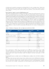
Table 1. Mediators of immune cells and adipocytes involved in NASH.
Cytokines and Adipocytokines Transcription Others
chemokines factors
Adiponectin Osteopontin
TNF-α Leptin NF-kB / IKKß SAA
IL-1α/ß Resistin JNK-1 CRP
Gp130 family (IL-6, CNTF) PBEF / Nampt / Visfatin PPARγ FABP-4
IL-10 RBP-4 SREBP-1c Oxidative stress
IL-18 IL-37 LXR ER stress
MCP-1 FXR iNOS
MIP-1α/ß Selectins
RANTES ICAM-1
VCAM-1
TLR-4/5
Key to abbreviations: CNTF, ciliary neurotrophic factor; FABP, fatty acid binding protein; ICAM,
intercellular adhesion molecule; iNOS, inducible nitric oxide synthase; LXR, liver X receptor; MCP,
monocyte chemoattractant protein; MIP, macrophage inflammatory protein; RBP, retinol binding
protein; SAA, serum amyloid A; VCAM, vascular cell adhesion molecule. See glossary for other
definitions.
Extrahepatic tissues as major sources of inflammatory mediators: key role for the adipose
tissue
The adipose tissue has been shown in many studies to reflect a major source of inflammatory mediators
in cases of obesity and related disorders. In severe human obesity visceral adipose tissue in particular,
but also subcutaneous adipose tissue constitute important sources of pro-inflammatory cytokines
such as IL-1β,, IL-6 or TNFα. Concentrations of all these mediators exceed their liver concentrations
The International Liver Congress™ 2015 • Vienna, Austria • April 22–23, 2015 85
especially experimental, studies suggest that microbiotal factors are driving forces of hepatic steatosis
and inflammation, involving certain toll-like receptors and induction of pro-inflammatory cytokines
such as TNFα.
Innate immune signals: crucial in NASH pathogenesis
The MetS is commonly observed in obesity and is thought to develop through the interaction of various
genetic and environmental factors. A complex and still poorly characterized interaction between the
intestinal microbiota and the innate system may be involved in metabolic dysfunction. MetS, diabetes and
obesity are characterized by low-grade inflammation, and adipocytokines play a central role. A profound
effect of the pattern recognition receptor TLR5 (activated by bacterial flagellin) on structural microbial
composition and its consequences for the pathogenesis of the MetS has recently been demonstrated
[7]. Mice lacking the TLR5 receptor exhibit hyperphagia and develop a MetS with hyperlipidemia,
hypertension, IR and obesity. Metabolic changes in TLR5 deficient mice resulted in abnormalities of the
intestinal microbiota. Transfer of this altered microbiota TLR5 deficient mice into gnotobiotic mice led
to MetS. These data provide not only experimental evidence that innate immune signalling is critical in
the development of MetS and a fatty liver, but also suggest that alterations in the intestinal microbiota
can be sufficient to induce the MetS and probably, crucially, drive the evolution of inflammation in
NASH.
Table 1. Mediators of immune cells and adipocytes involved in NASH.
Cytokines and Adipocytokines Transcription Others
chemokines factors
Adiponectin Osteopontin
TNF-α Leptin NF-kB / IKKß SAA
IL-1α/ß Resistin JNK-1 CRP
Gp130 family (IL-6, CNTF) PBEF / Nampt / Visfatin PPARγ FABP-4
IL-10 RBP-4 SREBP-1c Oxidative stress
IL-18 IL-37 LXR ER stress
MCP-1 FXR iNOS
MIP-1α/ß Selectins
RANTES ICAM-1
VCAM-1
TLR-4/5
Key to abbreviations: CNTF, ciliary neurotrophic factor; FABP, fatty acid binding protein; ICAM,
intercellular adhesion molecule; iNOS, inducible nitric oxide synthase; LXR, liver X receptor; MCP,
monocyte chemoattractant protein; MIP, macrophage inflammatory protein; RBP, retinol binding
protein; SAA, serum amyloid A; VCAM, vascular cell adhesion molecule. See glossary for other
definitions.
Extrahepatic tissues as major sources of inflammatory mediators: key role for the adipose
tissue
The adipose tissue has been shown in many studies to reflect a major source of inflammatory mediators
in cases of obesity and related disorders. In severe human obesity visceral adipose tissue in particular,
but also subcutaneous adipose tissue constitute important sources of pro-inflammatory cytokines
such as IL-1β,, IL-6 or TNFα. Concentrations of all these mediators exceed their liver concentrations
The International Liver Congress™ 2015 • Vienna, Austria • April 22–23, 2015 85