Page 96 - EASL POSTGRADUATE COURSE
P. 96
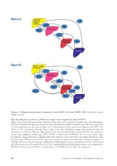
Figure 1A
Figure 1B
Figure 1. Fibrosis progression in patients with NAFL (1A) and NASH (1B). Taken from data in
Singh et al. [6].
Non-histological predictors of fibrosis progression in patients with NAFLD
Many of the paired biopsy studies discussed above have also examined whether any non-histological,
clinical or biochemical features can help in risk stratification of NAFLD patients into fibrosis progressors
vs. non-progressors. In the meta-analysis of 11 cohort studies [6] the presence of hypertension (OR 1.94,
95% CI 1.00–3.74) and a low AST/ALT ratio at the time of baseline biopsy was associated with the
progression of fibrosis. Features identified in some but not other studies (and therefore not significant
in the meta-analysis) include age, BMI, T2DM or MetS and HOMA-IR. In the most recent study
[7], fibrosis progressors had a significantly lower platelet count (P=0.04), and higher AST/ALT ratio
(P=0.04) and FIB-4 score (P=0.02) than non-progressors and a non-significantly higher prevalence of
T2DM (53% vs. 43%). At the time of follow-up biopsy, platelet count remained lower (P=0.0001) and
AST/ALT ratio (P=0.01) and FIB-4 (P=0.001) remained higher in progressors than in non-progressors.
NAFLD fibrosis score (P=0.001) and prevalence of T2DM was also higher in progressors.
96 Postgraduate Course Syllabus • Metabolic Liver Disease