Page 20 - EASL POSTGRADUATE COURSE
P. 20
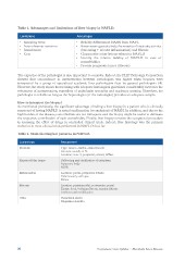
Table 1. Advantages and limitations of liver biopsy in NAFLD.
Limitations Advantages
• Sampling error • Reliably differentiate NASH from NAFL
• Inter-observer variation • Assess semi-quantitatively the severity of steatosis, activity
• Invasiveness
• Cost (balooning + lobular inflammation) and fibrosis
• Characterize other lesions related to NAFLD
• Identify the relative liability of NAFLD in case of
comorbidities
• Provide prognostic factor (fibrosis)
The expertise of the pathologist is also important to consider. Indeed, the FLIP Pathology Consortium
showed that concordance in interpretation between pathologists was higher when biopsies were
interpreted by a group of specialized academic liver pathologists than by general pathologists [4].
However, the study shows that training with adequate histological guidelines considerably increases the
robustness of interpretation, regardless of pathologist speciality and academic training. Therefore, the
pathologist is reliable as long as the hepatologist (or the radiologist) provides an adequate sample.
How to interpret the biopsy?
As mentioned previously, the significant advantage of taking a liver biopsy in a patient who is clinically
suspected of having NAFLD is actual confirmation (or exclusion) of NASH. In addition, and due to the
high burden of the disease, comorbidities are not infrequent and the biopsy might be useful to delineate
the respective contribution of each comorbidity. Finally, liver biopsy remains the recognized procedure
in assessing the effect of drugs in controlled clinical trials. Indeed, liver histology was the primary
endpoint in most clinical trials performed in NAFLD thus far.
Table 2. Main histological patterns in NAFLD.
Lesion type Assessment
Steatosis Type: macro-, medio-, microvacuole
Hepatocellular injury Amount: usually in %
Inflammation Location: zone 3, periportal, azonal, diffuse
Fibrosis
Other Ballooning and clarification of cytoplasm
Apoptotic body
MDB
Location: portal, periportal, lobular
Inflammatory cell type
Extent
Location: perisinusoidal, perivenular, portal
Extent: focal, bridging fibrosis, annular fibrosis
Architectural modification
Vacuolated nuclei
Megamitochondria
20 Postgraduate Course Syllabus • Metabolic Liver Disease