Page 21 - EASL POSTGRADUATE COURSE
P. 21
In NAFLD without comorbidities, the histopathological spectrum is relatively limited. Lesions should
be categorized into four main groups: steatosis, hepatocellular injury, inflammation and fibrosis (Table
2) [5]. Correct assessment is crucial for the characterization of the severity of changes that ultimately
lead to distinction between the processes considered to be non-progressive and not at risk of increased
liver disease mortality (i.e., NAFL) and those with features linked to progression of liver injury
(i.e., steatohepatitis, NASH). The diagnosis of steatohepatitis is based on the association of liver fat
(macrovacuolar or mediovesicular steatosis of ≥5%), hepatocyte ballooning and lobular inflammation
[6]. Perisinusoidal fibrosis is a useful and frequent diagnostic feature but not included formally in the
diagnostic criteria of steatohepatitis. In the early stages, the pattern of injury follows a centrilobular
accentuation, although, at later stages, the lobular architecture is mutilated and the zonal distribution
is no longer visible. Other histological features can be seen in steatohepatitis but are not necessary for
the diagnosis of NASH: perisinusoidal fibrosis, polymorphonuclear infiltrates, MDB, apoptotic bodies,
clear vacuolated nuclei, microvacuolar steatosis, megamitochondria and portal inflammation. Portal
inflammation is a frequent feature in pediatric NASH, but can be seen in adults and may be associated
with more severe disease. When steatosis is present but lobular inflammation or ballooning are absent,
the minimal requirements for steatohepatitis are not met, and the diagnosis should be NAFL (i.e. non-
NASH NAFLD). The terms ‘probable’ or ‘possible NASH’ should be abandoned because they create
confusion.
A final goal of liver biopsy in this setting is the semi-quantitative evaluation of the severity of injuries.
Indeed, although the dichotomized diagnostic approach (NAFL vs. NASH) is clinically useful, it is an
over-simplification that does not reflect the histological complexity of the disease. As with chronic liver
diseases, NAFLD might display a continuous spectrum of histological lesions so that splitting the disease
into two categories is useful but artificial. Therefore, semi-quantitative scoring system might better
reflect the complexity of the histological pattern. These scoring systems are currently of limited value in
common practice but are extremely useful in the context of clinical trials. The NASH Clinical Research
Network (NASH CRN) from the United States and the European FLIP Pathology Consortium have
both contributed towards an accurate histological evaluation of NAFLD. The NAS (NAFLD Activity
Score) described by the NASH CRN is the unweighted sum of steatosis (0 to 3), inflammation (0 to 3)
and ballooning (0 to 2) [7]. It is not designed to be a surrogate for the diagnosis of steatohepatitis but
rather a crude evaluation of the severity of the disease, once the diagnosis of NASH has been established
by the overall pathological assessment. Although the NAS is correlated with aminotransferase and
HOMA values, to date there is unfortunately no demonstration of any prognostic value of the NAS [8].
While most patients with a NAS <3 and a NAS >4 are bona fide NAFL and NASH, respectively, there
is a grey zone (NAS = 3 or 4) that includes both cases with NAFL and NASH. Consequently, its use as
a histological outcome in therapeutic trials is of questionable clinical relevance.
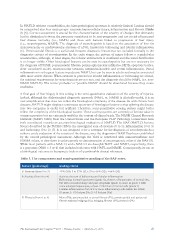
Table 3. The components and semi-quantitative grading of the SAF score.
Feature (grade range) Grading criteria
S: Steatosis (from 0 to 3) <5% (S0); 5 to 33% (S1); 33 to 66% (S2); >66% (S3)
A: Activity (from 0 to 4)
Activity: the sum of ballooning and lobular inflammation
F: Fibrosis (from 0 to 4) Ballooning: normal hepatocytes (grade 0), clusters of hepatocytes of normal size,
but with a rounded shape and pale cytoplasm (grade 1); same as grade 1 with
some enlarged hepatocytes, at least 2-fold that of normal cells (grade 2)
Lobular inflammation: foci of 2 or more inflammatory cells within the lobule
(0: none; 1: <2 foci per 20x; 2: >2 foci per 20x)
None (F0); perisinusoidal or portal fibrosis (F1); perisinusoidal and periportal
fibrosis without bridging (F2), bridging fibrosis (F3); cirrhosis (F4)
The International Liver Congress™ 2015 • Vienna, Austria • April 22–23, 2015 21
be categorized into four main groups: steatosis, hepatocellular injury, inflammation and fibrosis (Table
2) [5]. Correct assessment is crucial for the characterization of the severity of changes that ultimately
lead to distinction between the processes considered to be non-progressive and not at risk of increased
liver disease mortality (i.e., NAFL) and those with features linked to progression of liver injury
(i.e., steatohepatitis, NASH). The diagnosis of steatohepatitis is based on the association of liver fat
(macrovacuolar or mediovesicular steatosis of ≥5%), hepatocyte ballooning and lobular inflammation
[6]. Perisinusoidal fibrosis is a useful and frequent diagnostic feature but not included formally in the
diagnostic criteria of steatohepatitis. In the early stages, the pattern of injury follows a centrilobular
accentuation, although, at later stages, the lobular architecture is mutilated and the zonal distribution
is no longer visible. Other histological features can be seen in steatohepatitis but are not necessary for
the diagnosis of NASH: perisinusoidal fibrosis, polymorphonuclear infiltrates, MDB, apoptotic bodies,
clear vacuolated nuclei, microvacuolar steatosis, megamitochondria and portal inflammation. Portal
inflammation is a frequent feature in pediatric NASH, but can be seen in adults and may be associated
with more severe disease. When steatosis is present but lobular inflammation or ballooning are absent,
the minimal requirements for steatohepatitis are not met, and the diagnosis should be NAFL (i.e. non-
NASH NAFLD). The terms ‘probable’ or ‘possible NASH’ should be abandoned because they create
confusion.
A final goal of liver biopsy in this setting is the semi-quantitative evaluation of the severity of injuries.
Indeed, although the dichotomized diagnostic approach (NAFL vs. NASH) is clinically useful, it is an
over-simplification that does not reflect the histological complexity of the disease. As with chronic liver
diseases, NAFLD might display a continuous spectrum of histological lesions so that splitting the disease
into two categories is useful but artificial. Therefore, semi-quantitative scoring system might better
reflect the complexity of the histological pattern. These scoring systems are currently of limited value in
common practice but are extremely useful in the context of clinical trials. The NASH Clinical Research
Network (NASH CRN) from the United States and the European FLIP Pathology Consortium have
both contributed towards an accurate histological evaluation of NAFLD. The NAS (NAFLD Activity
Score) described by the NASH CRN is the unweighted sum of steatosis (0 to 3), inflammation (0 to 3)
and ballooning (0 to 2) [7]. It is not designed to be a surrogate for the diagnosis of steatohepatitis but
rather a crude evaluation of the severity of the disease, once the diagnosis of NASH has been established
by the overall pathological assessment. Although the NAS is correlated with aminotransferase and
HOMA values, to date there is unfortunately no demonstration of any prognostic value of the NAS [8].
While most patients with a NAS <3 and a NAS >4 are bona fide NAFL and NASH, respectively, there
is a grey zone (NAS = 3 or 4) that includes both cases with NAFL and NASH. Consequently, its use as
a histological outcome in therapeutic trials is of questionable clinical relevance.
Table 3. The components and semi-quantitative grading of the SAF score.
Feature (grade range) Grading criteria
S: Steatosis (from 0 to 3) <5% (S0); 5 to 33% (S1); 33 to 66% (S2); >66% (S3)
A: Activity (from 0 to 4)
Activity: the sum of ballooning and lobular inflammation
F: Fibrosis (from 0 to 4) Ballooning: normal hepatocytes (grade 0), clusters of hepatocytes of normal size,
but with a rounded shape and pale cytoplasm (grade 1); same as grade 1 with
some enlarged hepatocytes, at least 2-fold that of normal cells (grade 2)
Lobular inflammation: foci of 2 or more inflammatory cells within the lobule
(0: none; 1: <2 foci per 20x; 2: >2 foci per 20x)
None (F0); perisinusoidal or portal fibrosis (F1); perisinusoidal and periportal
fibrosis without bridging (F2), bridging fibrosis (F3); cirrhosis (F4)
The International Liver Congress™ 2015 • Vienna, Austria • April 22–23, 2015 21