Page 43 - EASL POSTGRADUATE COURSE
P. 43
Increased fat intake and a sedentary lifestyle can promote adiposopathy as well as visceral and ectopic
fat accumulation [6].
Visceral fat accumulation is an independent risk factor for the development of insulin resistance and for
hepatic fat accumulation. Lean subjects with NAFLD tend to have increased VAT [2, 6]. Independently
of obesity, VAT is associated with alterations in both glucose and lipid metabolism [4].
The presence of large hypertrophic adipocytes has been linked to ectopic fat deposition and increased
risk of metabolic dysfunction [5]. Ectopic fat in the liver (NAFLD) is frequently associated with hepatic
insulin resistance, excess VLDL secretion and decreased insulin clearance [4, 5]. Pancreatic fat is
associated with beta cell dysfunction and apoptosis. Intramyocellular triglycerides are associated with
impaired glucose metabolism and decreased mitochondrial function. Intramyocardial fat is associated
with impaired organ metabolism, increase oxidative stress and reduced mitochondrial function [4, 5].
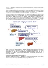
Figure 2. Lipotoxicity and progression to NASH. In the progression from steatosis to NAFLD,
NASH and cirrhosis it has been proposed that obesity, dyslipidemia, diabetes and excess
fat intake are the ‘first hit’ determining hepatic TG accumulation. A ‘second hit’, due to
lipotoxicity and oxidative stress, could be responsible for liver damage and progression to
NASH and cirrhosis.
Adipose tissue insulin resistance and lipotoxicity
Large adipocytes are more resistant to the antilipolytic effect of insulin, thus these subjects display
increased lipolysis and fatty acid overflow promoting ectopic fat and NAFLD.
The International Liver Congress™ 2015 • Vienna, Austria • April 22–23, 2015 43
fat accumulation [6].
Visceral fat accumulation is an independent risk factor for the development of insulin resistance and for
hepatic fat accumulation. Lean subjects with NAFLD tend to have increased VAT [2, 6]. Independently
of obesity, VAT is associated with alterations in both glucose and lipid metabolism [4].
The presence of large hypertrophic adipocytes has been linked to ectopic fat deposition and increased
risk of metabolic dysfunction [5]. Ectopic fat in the liver (NAFLD) is frequently associated with hepatic
insulin resistance, excess VLDL secretion and decreased insulin clearance [4, 5]. Pancreatic fat is
associated with beta cell dysfunction and apoptosis. Intramyocellular triglycerides are associated with
impaired glucose metabolism and decreased mitochondrial function. Intramyocardial fat is associated
with impaired organ metabolism, increase oxidative stress and reduced mitochondrial function [4, 5].
Figure 2. Lipotoxicity and progression to NASH. In the progression from steatosis to NAFLD,
NASH and cirrhosis it has been proposed that obesity, dyslipidemia, diabetes and excess
fat intake are the ‘first hit’ determining hepatic TG accumulation. A ‘second hit’, due to
lipotoxicity and oxidative stress, could be responsible for liver damage and progression to
NASH and cirrhosis.
Adipose tissue insulin resistance and lipotoxicity
Large adipocytes are more resistant to the antilipolytic effect of insulin, thus these subjects display
increased lipolysis and fatty acid overflow promoting ectopic fat and NAFLD.
The International Liver Congress™ 2015 • Vienna, Austria • April 22–23, 2015 43